

Medically reviewed by
USA Vein Clinics Medical Review Team
Vascular Specialists Focused on CVI Reviewed: November 2026
A vein ultrasound shows far more than what a physical exam can reveal. The test maps blood flow direction inside your leg veins, identifies damaged valves, measures whether blood is flowing backward, and can detect blood clots in both deep and superficial veins. For patients experiencing leg aching, swelling, visible varicose veins, or unexplained leg heaviness, the results of a vein ultrasound answer a central question: is there an underlying vein condition causing these symptoms, and how significant is it?
What a Vein Ultrasound Looks For?
A vein ultrasound uses duplex ultrasound technology to create real-time images of blood flow within your leg veins. Unlike an X-ray, it shows motion, allowing the specialist to evaluate how blood moves through each vein and whether the one-way valves are functioning properly.
By combining images of the veins with measurements of blood flow, the ultrasound helps identify venous reflux, blood pooling, and other circulation problems that may not be visible during a physical exam.
For more on the full evaluation process, see how varicose veins are diagnosed, including what to expect during your first appointment.
What a Vein Ultrasound Can Find?
By examining blood flow and the function of the vein valves, a vein ultrasound can reveal several important signs of vein disease. These are some of the most clinically significant findings.
Venous Reflux
Venous reflux is the most common finding in patients with vein disease. It occurs when the one-way valves inside the leg veins fail to close properly, allowing blood to flow backward toward the feet rather than upward toward the heart. On a duplex ultrasound, reflux appears as reversed blood flow during a brief breath-holding technique (a Valsalva maneuver) or when the patient shifts position. Reflux lasting more than 0.5 seconds in the deep veins, or more than 1 second in the superficial veins, is generally considered clinically significant.[1]
The location of the reflux and the amount of backward blood flow help the specialist determine whether vein disease is present and how much it may be contributing to your symptoms.
Chronic Venous Insufficiency (CVI)
Chronic venous insufficiency (CVI) is diagnosed when venous reflux is contributing to symptoms such as leg swelling, heaviness, pain, skin changes, or ulcers. The ultrasound helps confirm the diagnosis by showing abnormal blood flow and valve dysfunction within the affected veins. More extensive reflux often corresponds with more advanced vein disease and a greater likelihood of symptoms.
Varicose Vein Valve Incompetence
Varicose veins develop when superficial vein valves fail, and veins expand under increased internal pressure. A vein ultrasound identifies which specific veins are incompetent, how far the dysfunction extends along each vein, and whether the great saphenous vein, the small saphenous vein, or tributary veins are involved. This information is essential: treatment targets the incompetent vein directly, not just the visible bulge on the skin surface. Without knowing exactly which veins are affected and to what degree, targeted treatment cannot be planned accurately.
Deep Vein Thrombosis (DVT)
A vein ultrasound is the standard imaging test used to confirm or rule out a blood clot called deep vein thrombosis. The scan assesses whether blood is moving freely through the deep veins and whether the vessel wall is compressible. A healthy, clot-free vein collapses gently under the light pressure of the ultrasound probe. A vein containing a clot does not compress normally, and this compressibility test is one of the most reliable non-invasive indicators of DVT available.[2]
If DVT is suspected based on symptoms such as sudden leg swelling, warmth, or deep calf pain, a vein ultrasound is typically the first imaging step. DVT evaluation is usually performed in an emergency or urgent care setting and should not be delayed.
Superficial Thrombophlebitis
The scan can also detect clots that have formed inside superficial veins, most often inside varicose veins. This condition, called superficial thrombophlebitis, appears as a thickened, non-compressible segment within a superficial vein. One critical assessment the scan performs is determining whether the superficial clot is isolated or whether it has extended toward the junction with the deep venous system. That extension changes the clinical picture significantly and may change the management plan.
What Normal vs. Abnormal Results Look Like
A normal vein ultrasound shows blood flowing in the correct direction, vein valves that are working properly, and no evidence of blood clots or other abnormalities.
Abnormal results may show venous reflux, blood clots, damaged valves, or signs of chronic vein disease. Depending on the findings, the ultrasound can help explain symptoms such as leg pain, heaviness, swelling, skin changes, or nonhealing wounds.
The severity of abnormal findings can vary significantly. Some patients have only mild reflux in a small section of a vein, while others have more extensive disease affecting multiple veins.
For a broader overview of what an ultrasound for leg veins can reveal, see what a venous ultrasound can show.
Your vein specialist will review the results alongside your symptoms, medical history, and physical exam findings to determine whether treatment is needed.
What to Do Before a Vein Ultrasound
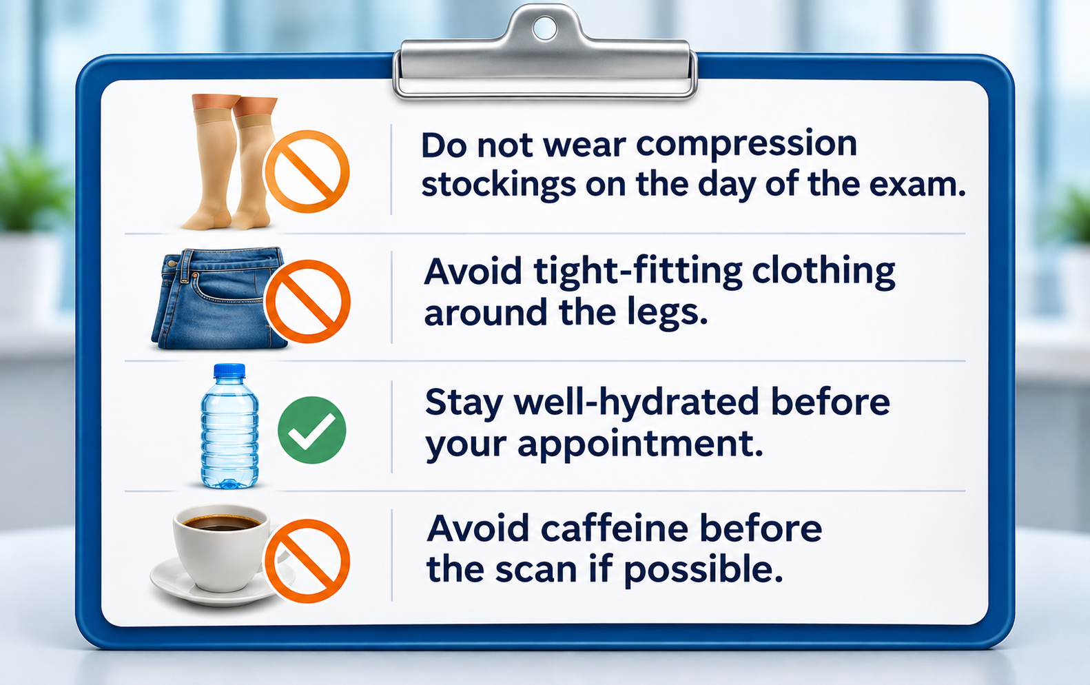
Preparation for a vein ultrasound is minimal, but a few things can affect image quality and scan accuracy:
- Do not wear compression stockings on the day of the exam. They alter blood flow dynamics temporarily and can affect reflux measurements.
- Avoid tight-fitting clothing around the legs. Loose pants or shorts make the scan easier and more comfortable.
- Stay well-hydrated before your appointment. Good hydration supports clear blood flow visualization on Doppler imaging.
- Avoid caffeine before the scan if possible. Caffeine can cause mild constriction in the vessels that may affect readings.
Beyond these steps, no fasting or special preparation is required. The scan uses no contrast agents, no radiation, and no needles. Most patients find it completely comfortable.
How Long It Takes to Get Results on a Duplex Vein Ultrasound?
At most vein clinics, including USA Vein Clinics, ultrasound results are reviewed in real time during the same appointment. A licensed vascular sonographer performs the scan while the vein specialist reviews the images as they are captured. By the end of your visit, your provider can typically walk you through what the scan showed, explain what the findings mean for your vein health, and outline potential next steps.
There is no waiting period for a lab to process images and issue a report. The information is available immediately after the scan is complete, which means your first appointment can include both the diagnostic step and an initial discussion about your options.
What Happens After a Vein Ultrasound
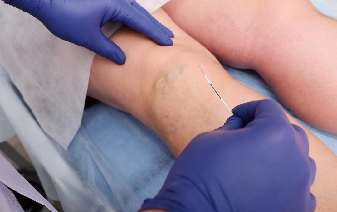
If the scan is normal, your vein specialist can reassure you that the symptoms you are experiencing do not appear to have a venous cause, and may recommend further evaluation for other contributing factors such as musculoskeletal or neurological causes.
If the scan reveals reflux, valve dysfunction, or other findings consistent with vein disease, your provider will discuss whether treatment may be appropriate based on the severity of the findings and the nature of your symptoms. Treatment recommendations are tailored to what the ultrasound shows specifically, which veins, how much reflux, and which anatomical pattern is driving your symptoms. No two patients have identical scan findings, which is why evaluation-first care matters.
If treatment is recommended, minimally invasive varicose vein treatment options at USA Vein Clinics include EVLT, RFA, VenaSeal, Varithena, and sclerotherapy, all performed in an outpatient setting. A follow-up ultrasound is typically scheduled after treatment to confirm the treated vein has closed properly.
Vein Evaluation
See What Your Veins Are Doing with a Duplex Vein Ultrasound
A vein ultrasound takes 30 to 60 minutes, requires no needles or radiation, and gives your vein specialist a complete picture of blood flow inside your legs.
If the scan finds venous reflux or valve dysfunction, your provider walks you through what it means and what, if anything, should be done. USA Vein Clinics offers outpatient evaluations at locations nationwide.
FAQs About Vein Ultrasounds
- Eklof B, et al. Revision of the CEAP classification for chronic venous disorders: Consensus statement. Journal of Vascular Surgery. 2004;40(6):1248-1252.
- Zierler BK. Ultrasonography and diagnosis of venous thromboembolism. Circulation. 2004;109(21 Suppl 1):I9-14.
- Cleveland Clinic. Duplex Ultrasound. my.clevelandclinic.org. Accessed November 2026.
Medical disclaimer: This article provides educational information about vein disease and diagnostic testing. It is not a diagnosis or medical advice. Only a qualified vein specialist can determine whether you have vein disease or recommend appropriate testing or treatment. If you have leg swelling, pain, or other vascular symptoms, consult a healthcare provider or contact USA Vein Clinics for an evaluation.


