
Medically reviewed by
USA Vein Clinics Medical Review Team
Vascular Surgeons and Vein Specialists
Reviewed: September 2026
May-Thurner syndrome (MTS), also known as iliac vein compression syndrome or Cockett syndrome, occurs when the right common iliac artery compresses the left common iliac vein against the lumbar spine. This vascular compression restricts blood flow from the left leg back to the heart, significantly elevating the risk of deep vein thrombosis (DVT). Because many individuals remain completely asymptomatic until persistent swelling or an acute blood clot develops, recognizing the clinical signs early is essential for preventing serious vascular complications.
May-Thurner syndrome is a frequently underdiagnosed anatomical cause of lower-extremity deep vein thrombosis (DVT), accounting for an estimated 2 to 5 percent of all lower-limb venous clotting events. While it predominantly affects women between the ages of 20 and 40, timely imaging and minimally invasive vein treatments allow this condition to be effectively managed on an outpatient basis.
Quick Facts: May-Thurner Syndrome
- Occurs when the right common iliac artery compresses the left common iliac vein against the fifth lumbar vertebra.[1]
- Accounts for approximately 2 to 5 percent of all lower-extremity deep vein blood clots.[1]
- Most frequently diagnosed in women aged 20 to 40, especially during or following pregnancy.[1]
- Outpatient treatment options like angioplasty and iliac vein stenting restore healthy blood flow without open surgery or hospital admission.
What Is May-Thurner Syndrome?
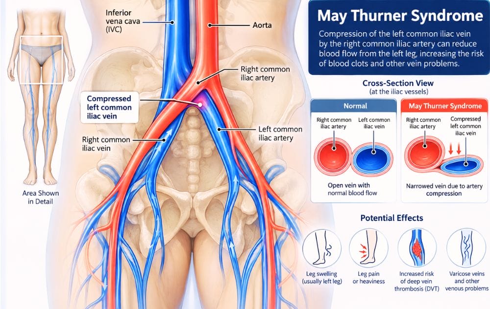
May-Thurner syndrome is a structural vascular condition localized in the pelvis. Arteries carry oxygen-rich blood away from the heart to the limbs under high pressure, while veins return deoxygenated blood back toward the heart under lower pressure. Where the circulatory pathways cross in the lower pelvis, the right common iliac artery lies directly over the left common iliac vein, crossing in front of the fifth lumbar vertebra ($L_5$).
Under continuous arterial pulsations, the rigid artery repeatedly compresses the softer vein against the underlying bone. Over time, this chronic mechanical irritation creates fibrous bands, spurs, or webs within the vein wall. These anatomical changes narrow the vessel lumen (stenosis), impede outflow from the left leg, and create an ideal environment for stagnant blood to clot.
How Rare Is May-Thurner Syndrome?
May-Thurner syndrome is far more prevalent than historical data suggested. Modern radiological studies reveal that mild to moderate left iliac vein compression is a common anatomical variant present in up to 20 to 30 percent of the general population. However, only a fraction of individuals develop symptomatic venous obstruction or clinical DVT.[1]
The condition is diagnosed 2 to 3 times more frequently in women than in men. Pregnancy represents a vulnerable period due to gestational blood volume increases, hormonal factors that increase vein wall elasticity, and direct mechanical pressure on pelvic veins from the growing uterus. Advances in cross-sectional imaging and catheter venography have made detection significantly more reliable in recent years.[1]
At USA Vein Clinics, our vascular specialists utilize non-invasive duplex ultrasound and specialized clinical protocols to evaluate pelvic vein compression and chronic venous outflow obstruction.
What Causes May-Thurner Syndrome and Increases DVT Risk?
The underlying cause of May-Thurner syndrome is anatomical compression of the left common iliac vein by the overlying right common iliac artery. While this mechanical pressure initiates the vessel narrowing, specific secondary risk factors often trigger the transition from an asymptomatic anatomical variation to an acute symptomatic condition or venous clot:
- Prolonged Immobilization: Long-haul flights, extended road travel, or post-surgical bed rest that slows systemic venous return.
- Pregnancy and Postpartum Period: Elevated circulating blood volume and direct pelvic compression compounded by pro-clotting hormonal shifts.
- Oral Contraceptives and Hormone Therapy: Estrogen-containing medications that increase hypercoagulability.
- Recent Surgical Procedures or Trauma: Orthopedic or pelvic operations that induce vascular inflammation or temporary immobility.
- Underlying Hypercoagulable Disorders: Inherited or acquired thrombophilias such as Factor V Leiden, prothrombin gene mutations, or antiphospholipid syndrome.
- Active Malignancy: Certain cancers and systemic chemotherapy treatments that alter blood viscosity and coagulation.
- Chronic Venous Disease: Pre-existing varicose veins or untreated chronic venous insufficiency (CVI).
May-Thurner Syndrome Symptoms
Because iliac vein compression can remain silent for years, it frequently goes undetected until blood flow becomes markedly restricted. When symptoms develop, they typically present exclusively in the left leg:
- Unilateral Left Leg Swelling: Noticeable fluid retention (edema) in the left thigh, calf, or ankle, often presenting with marked leg asymmetry.
- Persistent Aching and Heaviness: A dull, throbbing fullness in the left leg that worsens toward the end of the day or after prolonged standing.
- Venous Claudication: Severe cramping, tightness, or bursting pain in the thigh and calf brought on by physical exercise.
- Hyperpigmentation and Skin Changes: Reddish-brown skin discoloration (stasis dermatitis) or leathery skin texture around the lower calf and ankle.
- Venous Ulcers: Chronic, non-healing open wounds near the ankle bone in advanced cases of chronic outflow obstruction.
Vascular discomfort is frequently misattributed to muscle strain. Some patients also experience worsening leg pain at night, which should always be discussed during an in-depth clinical consultation.
When Symptoms Require Immediate Emergency Attention
The most urgent complication of May-Thurner syndrome is acute deep vein thrombosis. If a pelvic or deep leg clot dislodges, it can travel through the vena cava into the pulmonary arteries, resulting in a life-threatening pulmonary embolism (PE).
Warning Signs of Deep Vein Thrombosis (DVT):
- Sudden, rapid swelling in one leg (typically the left).
- Localized warmth and tenderness along the calf or inner thigh.
- Skin redness or bluish-purple discoloration.
- Severe, cramp-like tenderness when flexing the foot upward.
Warning Signs of Pulmonary Embolism (PE):
- Sudden, unexplained shortness of breath or rapid breathing.
- Sharp, stabbing chest pain that sharpens when inhaling deeply.
- Lightheadedness, dizziness, rapid heart rate, or fainting (syncope).
- Coughing up pink, frothy sputum or blood.
If you develop any symptoms of a pulmonary embolism, call 911 immediately or go to the nearest hospital emergency department.
How Is May-Thurner Syndrome Diagnosed?
Accurate diagnosis requires direct imaging of the pelvic venous anatomy because standard lower-leg ultrasound scans do not routinely visualize the iliac veins deep in the pelvis. A comprehensive diagnostic workup includes:
- Duplex Ultrasound: The initial non-invasive screening tool used to assess flow patterns, rule out lower-extremity DVT, and detect indirect signs of proximal outflow obstruction.
- CT Venography (CTV) or MR Venography (MRV): Cross-sectional imaging that clearly depicts the vascular crossing point and quantifies the degree of extrinsic compression between the artery and lumbar spine.
- Catheter Venogram and Intravascular Ultrasound (IVUS): The clinical gold standard. An interventional vascular specialist inserts a tiny ultrasound probe inside the vein to directly measure the cross-sectional area and pinpoint the location of intraluminal webs or spurs.
Treatment Options for May-Thurner Syndrome
The primary goals of May-Thurner syndrome treatment are relieving venous outflow obstruction, preventing initial or recurrent blood clots, and managing chronic swelling and skin changes. Clinical management is tailored according to whether an active clot is present.
1. Venous Angioplasty and Stenting (Non-Thrombotic Cases)
For patients with confirmed vein compression and chronic symptoms without an active clot, minimally invasive catheter-based treatment is the preferred standard. Under local anesthesia and light sedation, a vascular specialist performs:
- Balloon Angioplasty: A small balloon catheter is navigated into the narrowed left common iliac vein and briefly inflated to dilate the compressed segment.
- Iliac Vein Stenting: Because veins lack the muscular recoil of arteries, angioplasty alone is insufficient. A specialized self-expanding venous stent is deployed across the narrowed section. This medical-grade mesh scaffold withstands arterial pulsations, holds the vein open, and restores unhindered blood flow back to the heart.
2. Comprehensive Management for Active DVT
If May-Thurner syndrome has already triggered an extensive deep vein clot, treatment addresses both the acute thrombus and the underlying compression:
- Anticoagulation Therapy: Prescription blood thinners halt clot expansion and reduce the danger of pulmonary embolism.
- Catheter-Directed Thrombolysis / Thrombectomy: Specialized endovascular techniques break up or mechanically remove the clot, rapidly restoring lumen patency.
- Subsequent Stent Placement: Once the clot is cleared, an iliac stent is placed to prevent the vein from immediately re-clotting.
How USA Vein Clinics Can Help
USA Vein Clinics provides comprehensive, office-based vascular evaluations across our nationwide network of accredited outpatient centers. If you experience persistent left leg swelling, throbbing heaviness, or have a personal history of unexplained lower-extremity blood clots, our experienced team of vascular surgeons and vein specialists can conduct an in-depth diagnostic evaluation.
By combining high-resolution ultrasound imaging with personalized outpatient care plans, we help patients address the root cause of venous compression without hospital admissions or prolonged recovery windows. Learn more about the spectrum of vein disease we treat to understand how iliac vein compression interacts with overall circulatory health.
Next Step
Find Out If May-Thurner Syndrome Is Affecting Your Legs
Unexplained leg swelling or a personal history of blood clots warrants a focused vascular evaluation. Our expert vein specialists can determine whether iliac vein compression is contributing to your symptoms and map out a gentle, effective treatment path.
USA Vein Clinics offers minimally invasive, outpatient consultations covered by most major commercial insurance plans and Medicare.
Frequently Asked Questions About May-Thurner Syndrome
References
- Harbin MM, Lutsey PL. “May-Thurner syndrome: History of understanding and need for defining population prevalence.” Journal of Thrombosis and Haemostasis, 2020. National Center for Biotechnology Information (StatPearls).
Medical disclaimer: This educational article provides general clinical information regarding venous anatomy, May-Thurner syndrome, and outpatient treatment options. It is not intended as a substitute for professional medical diagnosis or individual treatment plans. Only an experienced vein specialist can diagnose vascular conditions through physical examination and diagnostic ultrasound. If you experience severe leg swelling, redness, sudden pain, or chest discomfort, seek immediate medical attention.